Chondroid Matrix X Ray

Pin By Phuong Hoai On Msk In 2020 Radiology Imaging Medical Medicine

Pin By Tnfri On Radiologi In 2020 Radiology Imaging Human Anatomy And Physiology Radiology

Pin By Phuong Hoai On Musculoskeletal Radiology Bone Diseases Pathology

Aneurysmal Bone Cyst Cysts Medical News First Aid Tips

Pin By محمد البدوي On Arthritis In 2020 Subtle Lockscreen Movie Posters
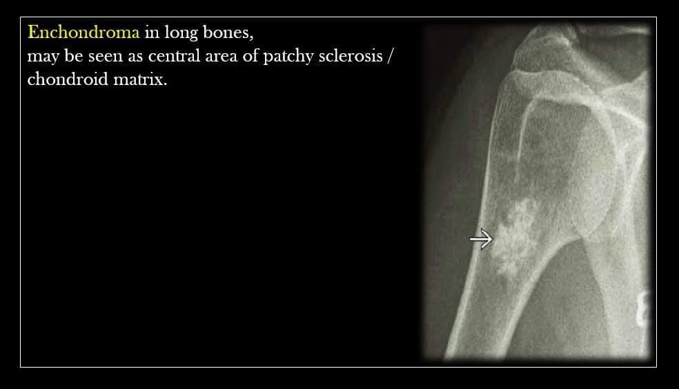
Pin By Raghav Tiwari On Msk In 2020 Central Area Arthritis Patchy
The surgical management of hand enchondroma without postcurettage void augmentation.
Chondroid matrix x ray.


Pin By Bassam Barca On Musculoskeletal Bone Diseases Sonography Pathology

Sclerotic Bone Tumors And Tumor Like Lesions Radiology Radiology Imaging Oral Pathology

Pin By Mauricio Zapata On Musculoskeletal Radiology Bone Diseases Pathology
Source : pinterest.com
